
Intro to 3D Cell Culture

Intro
Last week, we talked about cell-based assays. As a refresher, cell based assays typically involve growing cells in a monolayer, simulating a disease model (i.e. viral infection, mechanistic assays, etc), and testing your treatment on the disease model. In drug discovery, they are used before in vivo (animal) studies as a useful tool to find relevant molecules for your drug target and narrow down the optimal iteration of the treatment you’re testing(lead optimization). 3D cell culture offers an intermediary between cell based assays grown in a monolayer and in vivo animal studies. In this post, I’ll introduce 3D cell culture, compare it to 2D (monolayer) cell culture, and compare them to in vivo studies; furthermore, I’ll talk about organ on a chip, which is an advanced disease model making waves in the industry.
What is 3D Cell Culture?
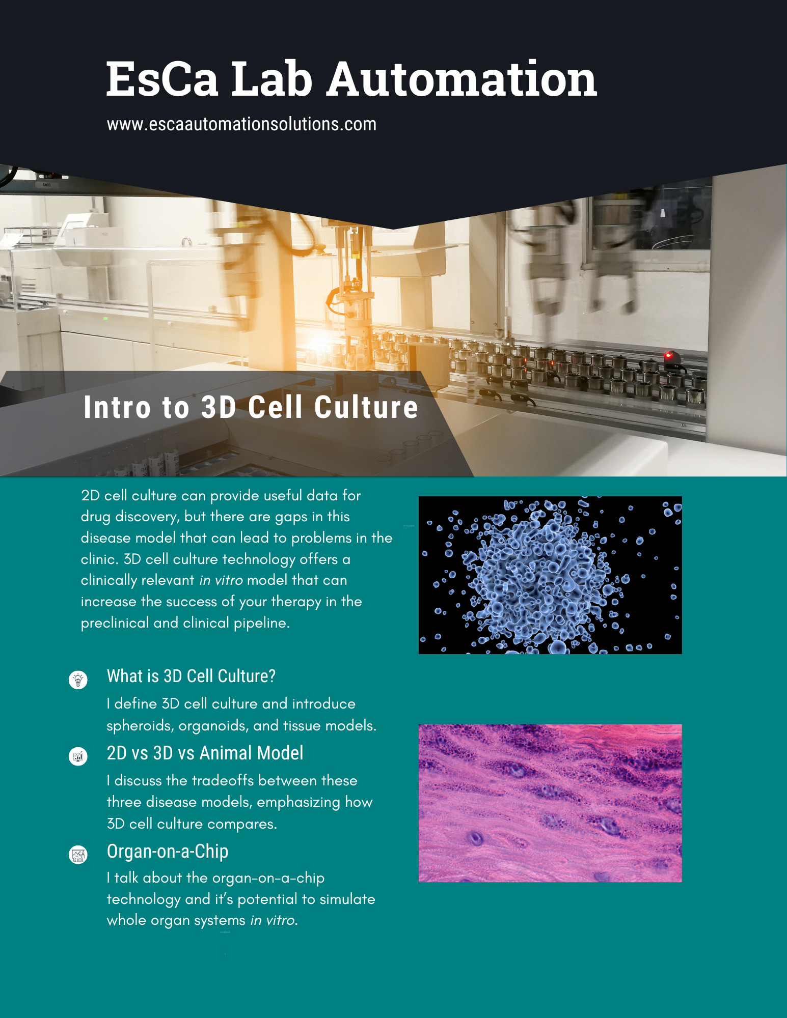
I had a really bad interview towards the start of my career. I thought 3D cell culture was another name for suspension cells since they technically spin around in a flask in 3 dimensions. I was wrong and didn’t get that job, so I’m here to make sure you don’t make the same mistake. 3D cell culture refers to cells cultured ‘adherently’ but in layers: the cells grow on top of each other. I say ‘adherently’ because there is often some sort of scaffolding involved: whether that’s a gel that solidifies to hold your spheroid in place, co-culture with fibroblasts, or microfluidic chips, to name a few. 3D cell culture can offer a more clinically relevant model than adherent cell culture because you can measure the different aspects of your drug in an environment where there is more interaction, like in the human body. There are three types of 3D cell culture that I’ll introduce to you here:
Spheroids are cells that grow in a spherical shape. They are arguably the simplestexample of 3D cell culture and can be grown in a matter of days. Spheroids are most effective in mimicking a tumor environment.Spheroids can be made with many different cell types with the right conditions.
Photo by National Cancer Institute on Unsplash Organoids are mini organs that are typically derived from stem cells. For drugs that target specific organs, this is the optimal model. An organoid could be made from patient-derived cells for personalized therapies that target things like neoantigens
Tissue Models like the air-liquid interface(ALI) can help mimic the skin, our largest organ. Not just the external epithelium, but also internally, like with the respiratory tract. They are composed of multiple cell types grown in layers and look qualitatively similar to actual human tissue under a microscope.
Photo by Pawel Czerwinski on Unsplash


Due to the complexity of developing these complex disease models, when sourcing organoids and tissue models, use a reputable company that uses rigorous characterization processes to validate the clinical relevance of their 3D cell culture rather than making them yourself. Again, it comes down to the tradeoff of whether you think it’s worth it to bring production of these technologies in house. But, it doesn’t hurt to do analysis to see if outsourcing the organoids will be cheaper than making them on-site.
3D vs 2D Cell Culture
2D cell-based assays are still necessary for drug discovery campaigns despite the availability of 3D models. Here are some limitations to consider when looking into 3D cell-based assays.
Cost 3D cell culture requires specialized consumables and techniques to make a relevant model. This extra lift may be cost consuming for earlier stages of drug discovery when you’re testing larger libraries in vitro.
Studies take longer because 3D cell cultures grow cells in layers. This makes any infection, treatment, and readout reagent take longer to penetrate the different layers and have an effect. If you’re trying to make these models in-house, differentiation and characterization can take weeks or even months before they’re ready for research purposes.
Imaging may not be possible with the most complex models. Firstly, the deeper you penetrate the layers of a 3D cell model, the more opaque your model is. Light is required for effective microscopy. Also, as I will discuss in a subsequent post, imaging more complex systems with high content imaging needs a ton of storage space and analysis.
3D cell culture technology is novel. This means that there will not be a ton of support for your research.
Your choice to use a 3D or 2D disease model depends on the question you’re trying to answer. One model isn’t inherently better than the other. Here are some things that a 3D model is best suited for:
Cytotox studies are more clinically relevant in 3D than in a monolayer because of the layers. You could see 100% death in a cell monolayer within a day, but that’s not what happens in the body.
More surface area in a 3D model can boost signal for less sensitive assays. If you engineer a cell line to express luciferase for a certain cellular mechanism, but the expression is low, consider growing them in a spheroid to boost the signal. Consider eplasia plates from Corning, which contain wells within each well of the plate for a spheroid to grow.
Co-culture is easier in 3D. If you want to see the interaction between different cell types, co culturing is an option; however, if you try to co-culture in a monolayer, faster-growing cells can outcompete other cell lines for space in each well. By growing them in layers, you can achieve the desired characteristics of your disease model more easily.
In the context of drug discovery, 3D cell culture models are not the industry standard. Remember that in drug discovery, you’re trying to narrow down the amount of material you’re testing because testing gets pricier as you proceed down the drug discovery pipeline. Right now, 3D cell-based assays fit somewhere in between 2D cell-based assays and in vivo studies. Let’s take a closer look at the comparison between 3D in vitro studies and in vivo studies.
3D Cell Culture Models and Animal Studies

3D cell culture can create clinically relevant models for disease when making drugs for humans. Animal models are important to see how drugs interact within a living organism, but these are not human. As of now, animal models are still required before testing anything in a clinical trial. One pervasive question that comes up when examining 3D cell culture is: will 3D cell culture models be able to replace in vivo studies any time soon? The short answer is no, but here are some advantages of 3D in vitro models over in vivo studies.
3D cell models can be automated. As I outlined in another post, stem cell culture can be automated as can the testing of the material in the assay. Differentiating, maintaining, and assaying your 3D cell culture can be done on robots.
There is more consistency in an in vitro model. When culturing cells, you can control many variables that lead to the desired characteristics for your disease model. This is especially true when you use automation, which increases the interoperability of protocols between users. You can’t control for animal behavior, like cannibalism between rodents, or automate steps in your protocol, like harvesting organs to isolate relevant cells.
Higher Throughput in 3D models means cheaper studies for further lead optimization. An in vivo scientist can only process so many animals.
3D cell culture is more ethical than animal studies. In an animal study, you’re subjecting animals to stress for an extended period only for them to be euthanized so their organs can be harvested and tested. This isn’t the case with cells.
But, as I said, in vitro studies do not capture the complexity of interactions within an organism. I would like to introduce an up and coming technology that attempts to do just that.
Organ-on-a-Chip

The name of this technology speaks for itself. Leveraging biology, microfluidics, engineering, and materials science, you can simulate organs on small chips. At about the size of a AA battery, this technology can be used to create clinically relevant disease models. Organs that have been simulated in this chip model include: lung , brain, heart, liver, kidney, skin, and gut.
This informative publication from the NIH details how this technology works. 3D cultured cell organoids are joined by channels within a chip. The channels allow for fluids and gasses to be exchanged within the system. This means nutrients can go in and waste can go out. Drugs can also be dosed in a gradient flow for more clinically relevant drug property data.
You can also have multiple organs on a chip! This brings us a step closer to testing drugs in an environment where you can measure the interaction of your drug and whole organ systems. As this technology advances, we will see more integration of high throughput automation: increasing the chance of a successful therapy in clinical trials by using the clinically relevant organ-on-a-chip system. Again, this is a complex technology, so unless you have an entire R&D department that can get organ-on-a-chip production up and running, buy them from a reputable source. Those will be properly characterized and ready to go.
Imagine a drug discovery workflow where the screening starts virtually, moves to 2D cell culture, then 3D cell culture before moving to in vivo animal studies. The preclinical pipeline of the future could drastically reduce drug discovery timelines and costs, especially when paired with AI drug discovery. Not to mention increasing the chance of your drug reaching the market!
As of now, we still need animal studies, but I’d like to imagine a world where we could skip those and obtain more clinically relevant data in vitro. At the end of the day, the human body is a complex system, and it’s hard to mimic all of the interactions that happen as of now.
Conclusion
3D cell culture offers a promising middle ground between traditional 2D cell-based assays and in vivo animal studies. While 3D cell culture presents challenges, it also provides numerous advantages, including increased clinical relevance and the potential for automation and higher throughput. As research in this area continues to advance, and with the development of innovative technologies like organ-on-a-chip, 3D cell culture is poised to play an increasingly important role in drug discovery and development, potentially leading to more efficient and effective treatments for a wide range of diseases.